Pauwels R, et al. Scatter-to-primary ratio in dentomaxillofacial cone-beam CT: effect of field of view and beam energy. Dentomaxillofac Radiol. 2021;50(8):20200597.
Article
PubMed
PubMed Central
Google Scholar
Jacobs R. Preoperative radiologic planning of implant surgery in compromised patients. Periodontol 2000. 2003;33(1):12–25.
Article
PubMed
Google Scholar
Jacobs R, et al. Cone beam computed tomography in implant dentistry: recommendations for clinical use. BMC Oral Health. 2018;18(1):88.
Article
PubMed
PubMed Central
Google Scholar
Pinheiro LR, et al. Effect of cone-beam computed tomography field of view and acquisition frame on the detection of chemically simulated peri-implant bone loss in vitro. J Periodontol. 2015;86(10):1159–65.
Article
PubMed
Google Scholar
Weiss R, Read-Fuller A. Cone beam computed tomography in oral and maxillofacial surgery: an evidence-based review. Dent J. 2019;7(2):52.
Article
Google Scholar
De Vos W, Casselman J, Swennen G. Cone-beam computerized tomography (CBCT) imaging of the oral and maxillofacial region: a systematic review of the literature. Int J Oral Maxillofac Surg. 2009;38(6):609–25.
Article
PubMed
Google Scholar
Molteni R. Prospects and challenges of rendering tissue density in Hounsfield units for cone beam computed tomography. Oral Surg Oral Med Oral Pathol Oral Radiol. 2013;116(1):105–19.
Article
PubMed
Google Scholar
Ruhrnschopf E-P, Klingenbeck K. A general framework and review of scatter correction methods in x-ray cone-beam computerized tomography. Part 1: scatter compensation approaches. Med Phys. 2011;38(7):4296–311.
Article
PubMed
Google Scholar
Altunbas C. Image corrections for scattered radiation. In: Shaw CC, editor. cone beam computed tomography. Boca Raton, FL: CRC Press; 2014. p. 129–47.
Google Scholar
Schulze R, et al. Artefacts in CBCT: a review. Dentomaxillofac Radiol. 2011;40(5):265–73.
Article
CAS
PubMed
PubMed Central
Google Scholar
Pauwels R, et al. Technical aspects of dental CBCT: state of the art. Dentomaxillofac Radiol. 2015;44(1):20140224.
Article
CAS
PubMed
Google Scholar
Kaasalainen T, et al. Dental cone beam CT: an updated review. Physica Med. 2021;88:193–217.
Article
Google Scholar
Cobos SF, et al. 3D-printed large-area focused grid for scatter reduction in cone-beam CT. Med Phys. 2023;50(1):240–58.
Article
PubMed
Google Scholar
Kim J, et al. Evaluation of a two-dimensional Moire-free antiscatter grid for cone-beam computed tomography. Med Phys. 2023;50(6):3435–44.
Article
CAS
PubMed
Google Scholar
Wiegert J, et al. Performance of standard fluoroscopy antiscatter grids in flat-detector-based cone-beam CT. Proc SPIE. 2004;5368:67–78.
Article
Google Scholar
Altunbas C, et al. Transmission characteristics of a two dimensional antiscatter grid prototype for CBCT. Med Phys. 2017;44(8):3952–64.
Article
CAS
PubMed
PubMed Central
Google Scholar
Fetterly KA, Schueler BA. Experimental evaluation of fiber-interspaced antiscatter grids for large patient imaging with digital x-ray systems. Phys Med Biol. 2007;52(16):4863–80.
Article
PubMed
Google Scholar
Singh V et al. Limitations of anti-scatter grids when used with high resolution image detectors. in Medical imaging 2014: physics of medical imaging. 2014. SPIE.
Kwan AL, Boone JM, Shah N. Evaluation of x-ray scatter properties in a dedicated cone-beam breast CT scanner. Med Phys. 2005;32(9):2967–75.
Article
PubMed
Google Scholar
Yu Z, Y Park, Altunbas C. Simultaneous scatter rejection and correction method using 2D antiscatter grids for CBCT. in Medical Imaging 2020: Physics of Medical Imaging. 2020. International Society for Optics and Photonics.
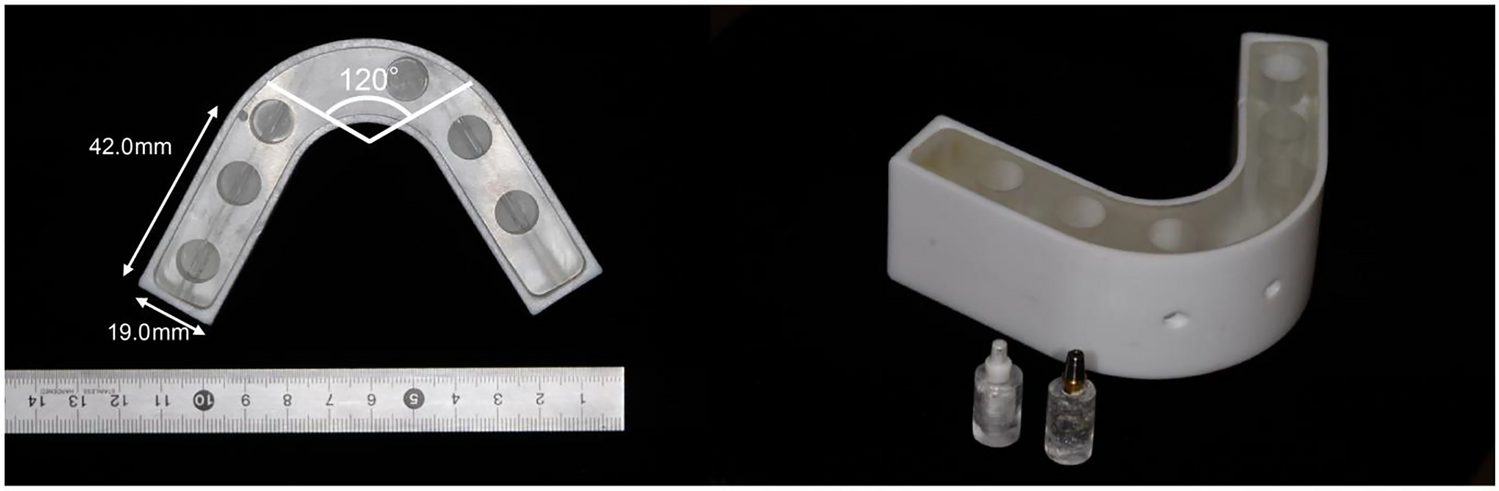
Park S-J, et al. Analysis of dimensions and shapes of maxillary and mandibular dental arch in Korean young adults. J Adv Prosthodontics. 2017;9(5):321–7.
Article
Google Scholar
Al-Zubair NM. The relationship between mandibular arch length and widths in a sample of Yemeni subjects with normal dento-Skeletal relationship. J Orthodontic Sci. 2013;2(4):120.
Article
Google Scholar
Welander U, et al. Standard forms of dentition and mandible for applications in rotational panoramic radiography. Dentomaxillofac Radiol. 1989;18(2):60–7.
Article
CAS
PubMed
Google Scholar
Gulliksrud K, Stokke C, Martinsen ACT. How to measure CT image quality: variations in CT-numbers, uniformity and low contrast resolution for a CT quality assurance phantom. Physica Med. 2014;30(4):521–6.
Article
Google Scholar
McCollough C, et al. Diagnostic reference levels from the ACR CT accreditation program. J Am Coll Radiol. 2011;8(11):795–803.
Article
PubMed
Google Scholar
Ruetters M, et al. Dental imaging using an ultra-high resolution photon-counting CT system. Sci Rep. 2022;12(1):7125.
Article
CAS
PubMed
PubMed Central
Google Scholar
Khader A, et al. Comparing radiation doses in CBCT and medical CT imaging for dental applications. J Pharmacy Bioallied Sci. 2024;16(Suppl 2):S1795–7.
Article
Google Scholar
Lee H, Badal A. A review of doses for dental imaging in 2010–2020 and development of a web dose calculator. Radiol Res Pract. 2021;2021(1):6924314.
PubMed
PubMed Central
Google Scholar
Deman P, et al. Dose measurements for dental cone-beam CT: a comparison with MSCT and panoramic imaging. Phys Med Biol. 2014;59(12):3201.
Article
CAS
PubMed
Google Scholar
Feldkamp L, Davis L, Kress J. Practical cone-beam algorithm. J Opt Soc Am A-Opt Image Sci Vision. 1984;1(6):612–9.
Article
Google Scholar
Biguri A, et al. TIGRE: a MATLAB-GPU toolbox for CBCT image reconstruction. Biomed Phys Eng Express. 2016;2(5): 055010.
Article
Google Scholar

Comments (0)