
Remember me

During this study period, 44 patients underwent transcatheter UHP-BA. Patient characteristics and diagnosis are shown in Table 1. A total of 78 UHP-BAs were performed on 57 lesions in 44 patients, with a median age of 6.6 years (range 104 days–37.2 years) and a median weight of 17.6 kg (3.0–66.4 kg). Of the 57 lesions, 42 required one procedure, 11 required two, 3 required three, and 1 lesion required five procedures. Among 44 patients, 13 had multiple lesions. Of these, 8 had bilateral PA lesions, 2 had unilateral PA lesions and Fontan fenestration lesions, 2 had unilateral PA lesions and shunt lesions, and 1 had unilateral PA lesions and superior vena cava (SVC) lesions. Of the 78 procedures, 74 were performed using the single-balloon technique, and 4 were performed using the double-balloon technique. Regarding the balloons used, in the single-balloon technique, the Yoroi® balloon was used in 51 procedures, and the Conquest® balloon was used in 23 procedures. In the double-balloon technique, two procedures used two Yoroi® balloons, another used two Conquest® balloons, and the remaining one used both a Yoroi® and a Conquest® balloon.
Table 1 Patient characteristics and diagnosisThe total success rate was 76%. The balloon-to-narrowest diameter ratio was 2.64 ± 0.69 in the success group. The balloon-to-narrowest diameter ratio was 1.92 ± 0.39 in the non-success group. The balloon-to-narrowest diameter ratio was significantly larger in the success group (p < 0.001).
The UHP-BA is for each target lesion described below (Table 2).
Table 2 Number of Patients, Lesions, and Procedures for Each Target SiteUHP-BA for PAFor PA lesions, a total of 39 UHP-BAs were performed on 32 lesions in 24 patients. The median age of the patients was 5.7 years (range 104 days–37.2 years), and the median weight was 15.7 kg (range 3.0–66.4 kg). Among the 24 patients, 16 had unilateral lesions, while 8 had bilateral lesions. Of the 32 lesions, 25 were treated with a single procedure, whereas 7 required two procedures. Of these, 3 UHP-BAs were performed at the stenting site (Fig. 1).
Fig. 1
Successful balloon angioplasty of left pulmonary artery stenosis (arrow) through a right ventricle pulmonary artery shunt (RVPA shunt). Angiography of RVPA shunt before (A) and after (B) ultra-high-pressure balloon angioplasty (4 mm × 20 mm Yoroi® balloon)
UHP-BA for all 39 PA lesions enlarged the stenotic lesion from 3.5 ± 1.1 mm to 5.3 ± 1.7 mm with 155 ± 26% improvement, decreased the pressure gradient across the stenotic lesion from 29 ± 24 mmHg to 23 ± 19 mmHg with 33 ± 36% improvement. The balloons used for PA were Yoroi® in 20 procedures and Conquest® in 16 procedures for the single-balloon technique. For the double-balloon technique, two procedures used two Yoroi® balloons, and another used a combination of Yoroi® and Conquest® balloons. The ratio of the balloon size to the narrowest diameter was 2.35 ± 0.50.
The procedural success rate for all PA lesions is 77% (30/39). In the PA lesions, the balloon-to-narrowest diameter ratio was 2.51 ± 0.45 in the success group and 1.81 ± 0.20 in the non-success group. The balloon-to-narrowest diameter ratio was significantly larger in the success group (p < 0.001).
UHP-BA for Fontan fenestrationFor Fontan fenestration lesions, 24 UHP-BAs were performed in 13 patients, with a median age of 12.9 years (4.7–16.5 years) and a median weight of 25.9 kg (13.1–48.4 kg). Of the 13 patients, 5 had stents implanted in the fenestration, and 8 had no stents. Of the eight patients without stents, 5 underwent one, 2 underwent two, and 1 underwent three procedures. Of the patients with implanted stents, 2 underwent one procedure, and one each underwent 2, 3, and 5 procedures. In total, 12 UHP-BAs were performed at the stenting site (Fig. 2).
Fig. 2
Successful balloon angioplasty of Fontan fenestration (arrow). Angiography of inferior vena cava before (A) and after (B) ultra-high-pressure balloon angioplasty (5 mm × 20 mm Yoroi® balloon)
UHP-BA for 24 Fontan fenestration lesions enlarged the stenotic lesion from 2.0 ± 0.6 mm to 3.7 ± 0.7 mm with 185 ± 59% improvement. Yoroi was used for all angioplasty. The ratio of the balloon size to the narrowest diameter was 2.86 ± 0.87.
The procedural success rate for all Fontan fenestration lesions is 75% (18/24). In the Fontan fenestration lesions, the balloon-to-narrowest diameter ratio was 3.13 ± 0.86 in the success group and 2.11 ± 0.25 in the non-success group. The balloon-to-narrowest diameter ratio was significantly larger in the success group (p < 0.01).
UHP-BA for SVCFor SVC lesions, 8 UHP-BAs were performed in 6 patients, with a median age of 4.3 years (1.4–9.0 years) and a median weight of 14.8 kg (9.4–24.3 kg). Four of the six patients had stenosis at the SVC-PA anastomosis after the bidirectional Glenn, and two had stenosis at the SVC-RA junction after biventricular repair. One of these patients had stents implanted. UHP-BA for SVC lesions enlarged the stenotic lesion from 4.0 ± 1.2 mm to 6.0 ± 1.5 mm with 152 ± 18% improvement, decreased the pressure gradient across the stenotic lesion from 3 ± 2 mmHg to 2 ± 2 mmHg with 69 ± 39% improvement. The procedural success rate for SVC lesions is 88% (7/8). The balloon used for SVC was Yoroi® in 1 and Conquest® in 7. The ratio of the balloon size to the narrowest diameter was 2.36 ± 0.63.
UHP-BA for shuntFor shunt lesions, 4 UHP-BAs were performed in 3 patients, with a median age of 3.5 years (2.3–5.1 years) and a median weight of 11.4 kg (6.6–13.0 kg). One of the three patients had a Blalock–Taussig shunt, one had a central aortopulmonary shunt, and one had a right ventricle-to-pulmonary artery shunt. None of these patients had stents implanted. Duration from shunt creation was 2 years (1–4.9 years). UHP-BA for shunt lesions enlarged the stenotic lesion from 2.1 ± 0.5 mm to 3.1 ± 0.4 mm with 150 ± 21% improvement. The procedural success rate for shunt lesions is 75% (3/4). Yoroi® was used for all angioplasty.
UHP-BA for other lesionsUHP-BA was performed for PA banding lesions and Fontan conduit lesions. For PA banding lesions, two UHP-BAs were performed on two lesions in two patients. Yoroi® was used for all angioplasty. UHP-BA for PA banding lesions enlarged the stenotic lesion from 2.8 and 2.8 mm to 3.0 and 3.1 mm, with 107 and 111% improvement, respectively. None of these procedures were considered procedural success. The ratio of the balloon size to the narrowest diameter was 2.5.
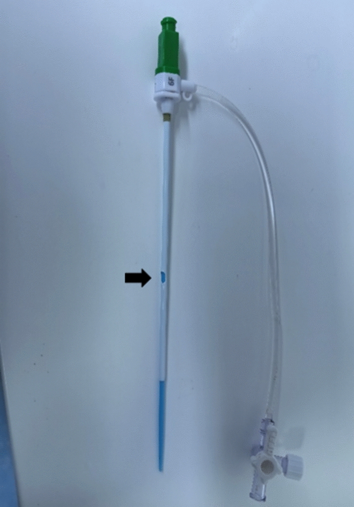
For Fontan conduit lesions, only one UHP-BA was performed using the double-balloon technique by two Conquest® balloons (Fig. 3). Although the stenotic lesion enlarged with 138% improvement, the pressure gradient did not reduce, and the procedure was not considered a procedural success.
Fig. 3
Balloon angioplasty of Fontan conduit (extra cardiac conduit with 20 mm PTFE). Angiography of inferior vena cava before (A) and after (C) ultra-high-pressure balloon angioplasty. B Double-balloon technique using 12 mm × 40 mm Conquest® balloon and 10 mm × 40 mm Conquest® balloon. Arrow: calcified conduit
Cases of upgrading from conventional balloon to UHP balloonAmong the 78 procedures, 11 required upgrading the balloon from a conventional balloon to a UHP balloon during the same session due to residual balloon waist. The target lesions included PA (7 procedures), fenestration (3 procedures), and SVC (1 procedure). The balloon was upgraded to a UHP balloon, but its diameter was not identical to that of the previously used conventional balloon and varied across cases. In all procedures upgraded to UHP-BA, the waist successfully disappeared. The diameter of the UHP balloon without a waist was significantly larger than the waist diameter observed with the conventional balloon (p < 0.01). UHP-BA for 11 upgrading lesions enlarged the stenotic lesion from 2.8 ± 1.2 mm to 5.0 ± 1.7 mm with 186 ± 90% improvement. The technical success rate was 91% (10/11). The only case not considered a technical success was a right PA stenotic lesion following arterial switch operation for complete transposition of the great arteries. Balloon angioplasty was first performed using a 10-mm conventional balloon, followed by UHP-BA with a Conquest balloon of the same diameter. As a result, the narrowest lesion improved from 2.9 mm to 4.0 mm; however, the improvement rate was limited to 145%, and the pressure gradient reduction did not meet the criteria.
ComplicationA complication of UHP-BA occurred in 3 of 78 UHP-BAs (3.8%). Two of the three patients had pulmonary hemorrhage, which was caused by guidewire injury, and they were treated with mechanical ventilation using positive end-expiratory pressure and recovered with conservative treatment. The remaining patient, who had undergone UHP-BA for SVC-PA anastomosis lesion, had pulmonary artery embolization due to migration of a thrombus at the stenotic lesion and required surgical thrombectomy. There was no perioperative mortality.
Follow-upThe median follow-up period was 3 years (range 1.6–4.5 years). During the follow-up period, additional treatment was performed on 35 of the 57 lesions. The reintervention rate was 61%. Additional balloon angioplasty (regardless of whether a UHP balloon was used) was performed on 31 lesions, while unplanned surgical intervention was selected for 4 lesions. The balloon-to-narrowest diameter ratio in patients with restenosis was 2.59 ± 0.72, while the balloon-to-narrowest diameter ratio in patients without restenosis was 2.38 ± 0.50, tending to be larger in patients with restenosis, but the difference was not significant.
Comments (0)