
Remember me


The study was conducted at the Tissue Culture Laboratory of the East Mediterranean Transitional Zone Agricultural Research Institute (EMTZARI). The acclimatization and transfer of plants obtained from in vitro culture to ex vitro conditions were performed in greenhouses belonging to EMTZARI.
Plant MaterialThe plant material used in this study consisted of the promising sumac genotype GN-61, obtained through selection breeding from the eastern Mediterranean region of Turkey. Specifically, this genotype was sourced from the Kahramanmaraş/Önsen district (N37°31.274′ E36°47.761′, altitude 669 m). Among the approximately 300 genotypes evaluated between 2019 and 2021, this genotype demonstrated the most favorable agronomic and botanical traits, including superior adaptability, productivity, and bioactive compound content (Adali et al. 2023). It was identified as the most promising candidate for clonal propagation studies due to its potential for large-scale cultivation and its suitability for meeting the growing demand in the medicinal and aromatic plant sector. Table 1 presents the location details of the GN-61 plant, along with the average values of various characteristics examined in 2020, 2021, and 2022. Explants were collected from 1-yr-old shoots of plants growing in the natural flora of the Kahramanmaraş-Önsen area during the early growing season (May 2022).
Table 1. Location information of the GN-61 plant and averages of the characteristics examined in 2020, 2021, and 2022 Culture Initiation, Surface Sterilization of Shoots, Culture Conditions, and Medium ComponentsExplants were obtained from young shoots approximately 20 cm in length. Fresh shoots were cut under field conditions and transported to the laboratory. The shoots were wrapped in moist paper towels, covered with aluminum foil, and stored at + 4 °C for 24 h. In the laboratory, the leaf blades were removed while the petioles remained intact, producing starting materials approximately 5 cm in length, each containing at least one node.
The axillary buds were rinsed under running tap water for 1 min and then dipped in 70% ethanol (v/v) for 3 s. Subsequently, the explants were transferred to a sterile cabinet and immersed in a 20% commercial bleach solution (Bingo, Istanbul Turkey) containing approximately 5% NaOCl with intermittent agitation for 20 min. After surface sterilization, the explants were rinsed three times with sterile distilled water for 5 min each.
The browned ends of explants that came into contact with NaOCl were trimmed slightly. Explants were transferred to the SPM medium, free of plant growth regulators (PGRs). Cultures were maintained at 25 ± 2 °C with a 16/8-h light/dark photoperiod using fluorescent lamps (Philips, Eindhoven, Netherlands), providing a photon flux density of 40 μmol∙m−2∙s−1.
Micro axillary buds from well-developed GN-61 sumac plants, cultivated on PGR-free SPM medium developed in Adali’s project (Adali 2023), were used in the experiments. The SPM medium was specifically designed as an alternative to the commonly used MS medium to optimize propagation conditions for shrub and woody plant species. The key differences between SPM and MS media lie in their macronutrient and micronutrient compositions, with SPM containing higher levels of NH₄NO₃, Ca(NO₃)₂, and specific micronutrients tailored to the nutritional requirements of shrub species. The SPM medium was supplemented with 4% (w/v) sucrose and 8 g L⁻1 agar (Table S1). The pH was adjusted to 5.7 using NaOH (Sigma-Aldrich, St. Louis, MO) before autoclaving at 121 °C and 1.2 bars for 20 min.
Micropropagation Stage in a Plantform Bioreactor System and Semi-solid Medium, and All MeasurementsIn the initial phase of comparing two propagation systems—semi-solid medium in glass jars and liquid medium in the TIS bioreactor (Plantform system, Plant form, Hjarup, Sweeden)—50 explants from each culture were placed in PGR-free SPM medium for proliferation. The newly developed shoots, measuring 2–3 cm in length and obtained from both media, were the starting material for the shoot proliferation experiments. The medium formulation for both liquid and semi-solid conditions was identical to that described in Table S1, with the only difference being the addition of 8 g L⁻1 agar to the semi-solid medium in the glass jars. The medium pH was adjusted to 5.7 in all cases. Each culture vessel in Plantform BIO contained 500 mL of medium. After 3 wk, the medium in the bioreactor was replaced with a fresh medium of the same composition. In contrast, the plantlets in the semi-solid medium were transferred to the fresh medium.
During the proliferation stage, 550-mL transparent glass bottles with lids were used as culture vessels for the semi-solid medium, each containing 65 mL of the nutrient medium. Fifty explants were placed per bioreactor and five per bottle, with three replicates per treatment.
This study aimed to simultaneously compare the effects of different treatments on axillary shoot formation in sumac plants using the semi-solid medium and TIS bioreactor (Plantform system) systems. The treatments involved supplementing the SPM medium with various cytokinins (1 mg L⁻1 BAP, TDZ, KIN, and ZEA) combined with the auxin IAA at a concentration of 0.25 mg L⁻1. IAA was specifically selected for its well-documented role in enhancing cell division and elongation in shoot proliferation studies. Furthermore, its compatibility with cytokinins makes it an ideal choice for optimizing proliferation without promoting excessive rooting (Lam et al., 2020; Etesami and Glick, 2024). All hormones were obtained from Duchefa Biochemie (Haarlem, Netherlands). The plants were cultured for 6 wk. In the Plantform BIO, the immersion frequency was set to 10 min every 8 h, and aeration was provided for 15 min every 6 h.
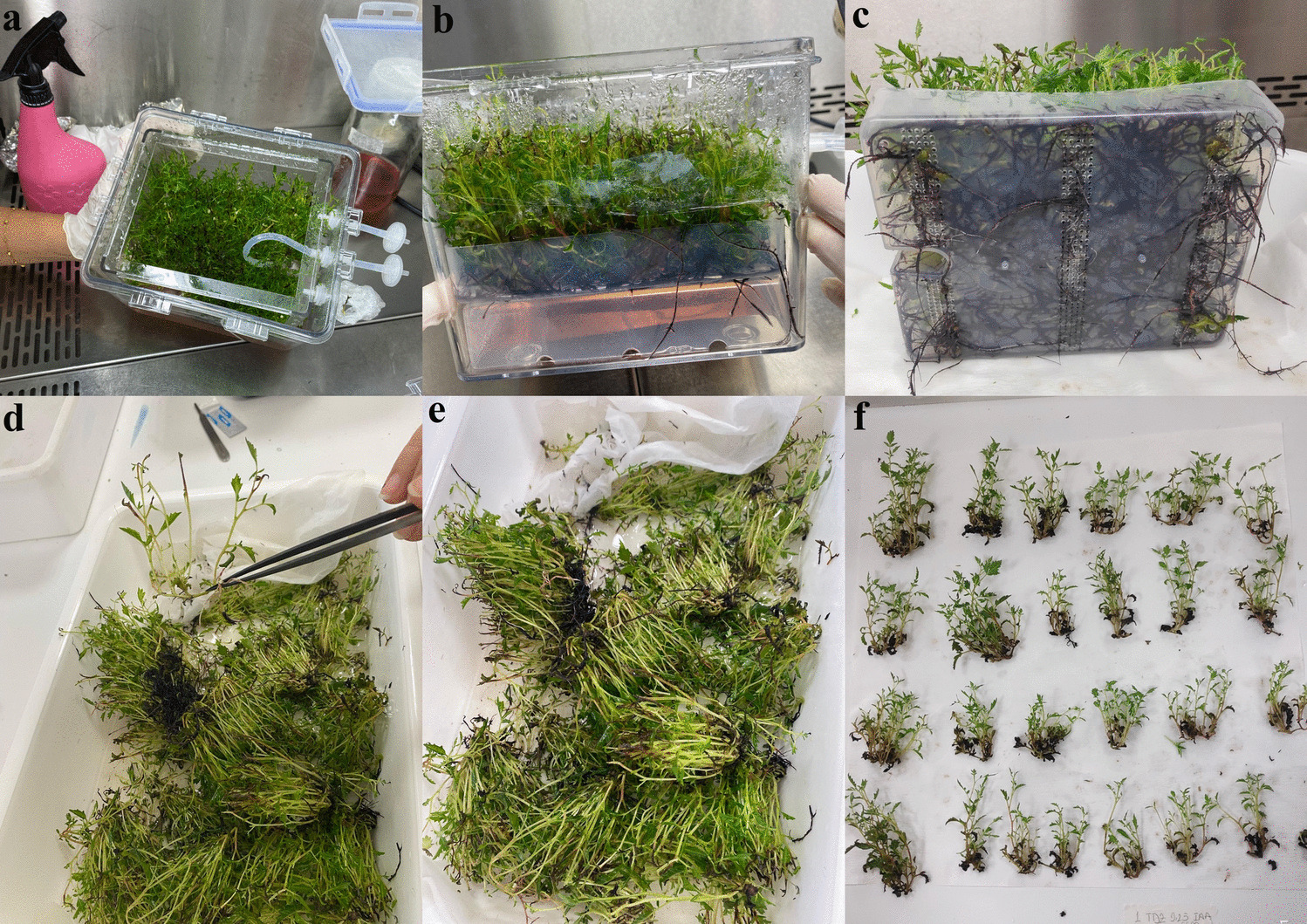
There was no extra rooting stage included in this study. Plantlets were directly evaluated after being collected from the respective systems, and no rooting medium or specific rooting treatments were applied. Shoot clusters from semi-solid medium bottles were carefully transferred using forceps to evaluate shoot proliferation. In the Plantform bioreactor, the entire plant community in the tray reservoir was transferred at once. Individual shoots were then separated from clusters using a scalpel and forceps. The number of shoots per explant, rooting percentage (%), shoot and leaf length (mm), leaf width (mm), and fresh and dry weight (g) were determined for each culture vessel. The sequential stages of micropropagation in the Plantform bioreactor system are illustrated in Fig. 1, showcasing the production process and the preparation of shoots for further analysis.
Figure 1.
Micropropagation stages of sumac (Rhus coriaria L.) in the Plantform bioreactor system. (a) General view of bioreactor system. (b) Shoot proliferation in the bioreactor. (c) Root structures developed in the bioreactor. (d) Separation of shoot clusters after culture. (e) Image after separation process. (f) Individual shoots prepared for further analysis.
For each treatment with different PGRs, the shoots developed from 15 explants in the two systems were weighed, including any newly formed calluses at the base of the shoots, using a precision balance to determine the average weight per explant. The fresh samples were dried in an oven at 45 °C for 48 h and weighed using a precision balance to determine the dry weight per explant.
Statistical AnalysesThe experiment followed a completely randomized design with three replicates per treatment. Each replicate comprised 15 explants from the semi-solid medium and Plantform BIO. Data were collected for several parameters, including the number of shoots per explant, rooting percentage, shoot length, average fresh and dry weight, shoot and leaf lengths, and leaf width. These data were statistically analyzed to determine significant differences between treatments. An analysis of variance (ANOVA) was performed to evaluate the effects of different plant growth regulator (PGR) treatments and culture systems on the measured parameters. Differences among means were determined using Tukey’s HSD (honestly significant difference) test at a significance level of p < 0.05.
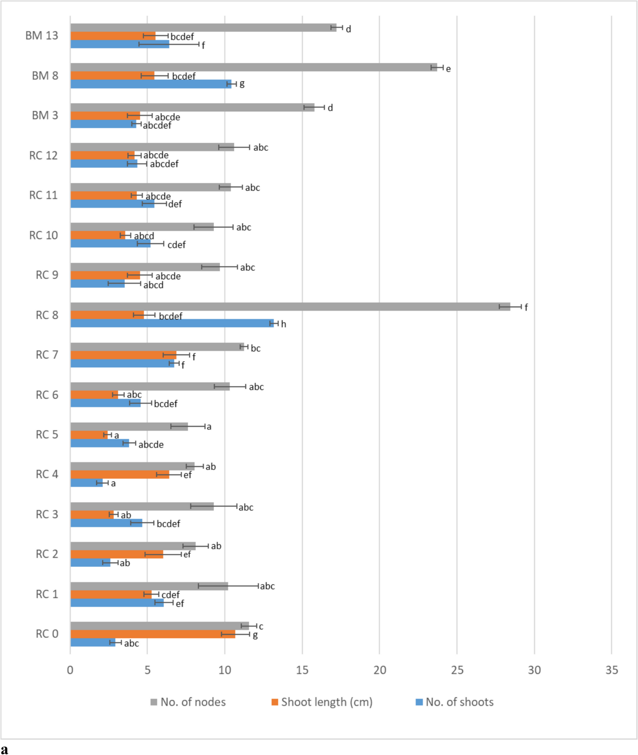
In cases where the data did not meet the assumptions of normality and homogeneity of variances, appropriate data transformations (e.g., log or square root transformation) were applied before conducting ANOVA. Descriptive statistics, including means and standard errors, were calculated for each treatment. Principal component analysis (PCA) was performed using JUMP 13.2 software (SAS Institute Inc., Cary, NC) to explore the interrelations between treatments and growth parameters. The results were visually represented in a biplot highlighting treatment groupings and parameter associations. The shoot proliferation rates and shoot lengths data were also presented in bar graphs to depict the differences among treatments and culture systems visually.
Comments (0)