2.1 Pan-cancer expression and functional analysis of ANAPC10
This study employed Sento Academic Online tools to assess the mRNA expression of ANAPC10 across various cancers and normal tissues. The BioWinford Platform was used to perform pan-cancer GSEA for the functional analysis of ANAPC10. These platforms leverage extensive data from the TCGA and GTEx projects and integrate them for more advanced analyses.
2.2 Analysis of ANAPC10 expression and its clinical correlations in OSCC
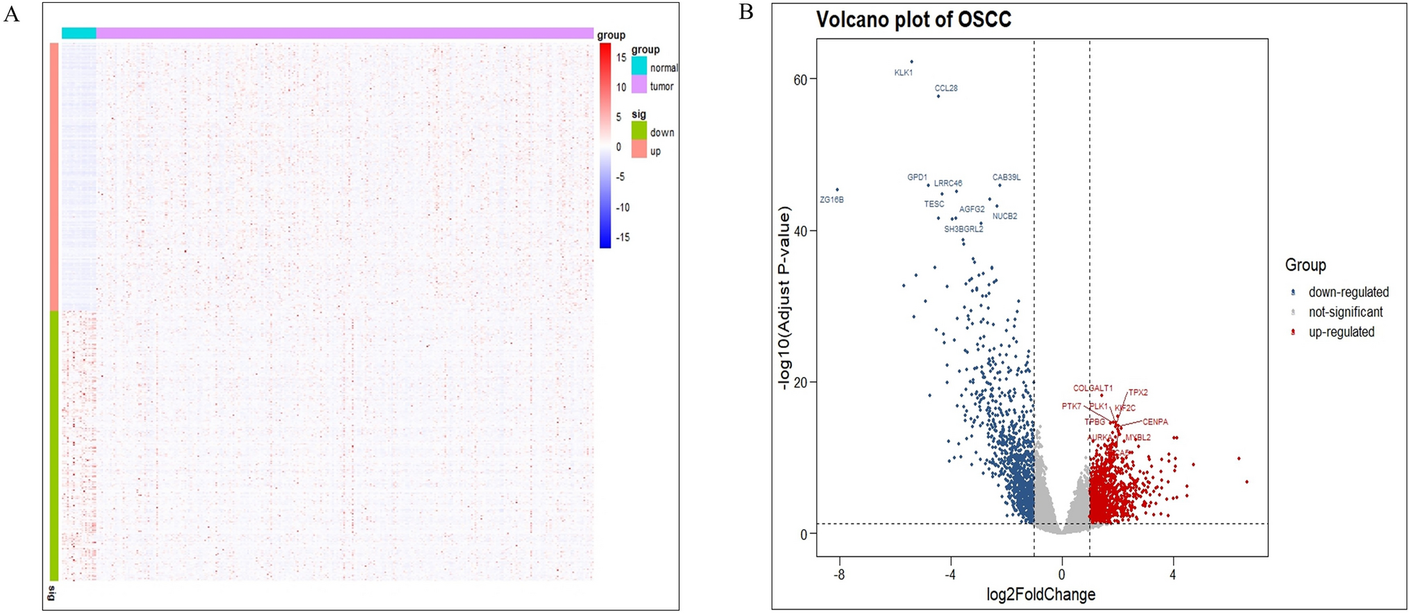
This study analyzed ANAPC10 expression in OSCC and normal oral epithelial cells based on paired and unpaired samples using the Sento Academic Online tool. Additionally, we analyzed variations in ANAPC10 expression across different clinical and pathological stages, primary therapy outcomes, histologic grades, cancerous sites, and sexes in patients with OSCC. We also assessed ROC curves to evaluate the diagnostic potential of ANAPC10 expression in OSCC. These platforms integrate and leverage extensive data from TCGA and GTEx projects for more advanced analyses.
2.3 Prognostic analysis and modeling of ANAPC10 in OSCC
In this study, we used the Sento Academic Online Tool to investigate the relationship between ANAPC10 expression and patient outcomes, including overall survival (OS), disease-specific survival (DSS), and progression-free interval (PFI), using data from TCGA and GTEx projects. We also created a prognostic model that included ANAPC10 expression as a Cox factor based on OSCC data from TCGA and evaluated its accuracy using a calibration curve.
2.4 Protein–protein interaction network construction and functional enrichment analysis
GeneMANIA was used to identify proteins interacting with ANAPC10 and to construct protein–protein interaction networks. Functional enrichment analysis of ANAPC10-associated genes was conducted using Gene Ontology (GO) and Kyoto Encyclopedia of Genes and Genomes (KEGG) pathways and Sento Academic online tools. We also examined the differential expression of these key genes in OSCC and adjacent noncancerous tissues.
2.5 Cell culture
Normal oral epithelial cells (HCP-H203) and various OSCC cell lines (ACC2, ACC3, SACC83, MEC1, and SCC9) were obtained from the Shanghai Cell Bank of the Chinese Academy of Sciences. Cells were cultured in DMEM, F12K, or RPMI 1640 complete medium supplemented with 10% fetal bovine serum (FBS) and 1% penicillin–streptomycin. Cells were maintained at 37 °C in a 5% CO2 atmosphere with regular medium changes to ensure optimal growth.
2.6 Design, synthesis, and transfection of small interfering RNAs
To study the function of ANAPC10, we designed and synthesized several small interfering RNA (siRNA) sequences, including a negative control (si-NC) and two specific siRNAs targeting ANAPC10. The negative control sequences are: 5'-UUCUCCGAACGUGUCACGUTT-3' and 5'-ACGUGACACGUUCGGAGAATT-3'. The ANAPC10-targeting siRNAs are: sequence 1, 5'-GCCACAGUCCUCAAGUUUUTT-3' and 5'-AAACUUGAGGACUGUGGCTTT-3'; sequence 2, 5'-GCCUCAUGGAUGAUGUUCUTT-3' and 5'-AAGACAUCAUCCAUAGGCCTT-3'. siRNAs were synthesized by Integrated DNA Technologies (IDT). Lipofectamine 3000 Transfection Reagent (Invitrogen, Cat# L3000008) was used to transfect the siRNAs into SCC9 cells at a concentration of 100 nM, following the manufacturer’s protocol, for 24 h. After transfection, cells were cultured for an additional 48 h to allow for effective siRNA expression.
2.7 RT-qPCR
Total RNA was extracted from the cells using TRIzol Reagent (Cat#15596018CN, Thermo, USA). The purity and concentration of the RNA were assessed using a spectrophotometer, and the concentrations were adjusted with DEPC-treated water. Qualified RNA was reverse-transcribed into cDNA using a specific kit, and ANAPC10 expression was quantified using GAPDH as an internal control. qRT-PCR was performed under the following conditions: initial denaturation for 10 min at 95 °C, followed by 40 cycles of 15 s at 95 °C and 1 min at 60 °C, with three technical replicates for each sample. The primers for ANAPC10, synthesized by Shanghai Shenggong Biological Engineering Co., Ltd., were: Forward primer: 5'-GCCACAGTCCGGAAGTCACT-3'; Reverse primer: 5'-TCACCCACTTGAGCTTGAGG-3'.
2.8 CCK-8 experiment
SCC9 cells transfected with si-ANAPC10 were seeded into 96-well plates at a density of 5000 cells per well. After 24 h of incubation, cell viability was assessed using the CCK-8 assay. The absorbance at 460 nm was measured to evaluate cell proliferation. The CCK-8 assay kit was purchased from Dojindo Molecular Technologies, Inc. (Japan, Cat# CK04).
2.9 Western blot analysis
Cells were lysed with RIPA buffer (Thermo, USA, Cat# 89900) containing protease and phosphatase inhibitors (Thermo, USA, Cat# 78440), then centrifuged at 14,000 rpm for 10 min at 4 °C to collect the protein-containing supernatant. The protein concentration was measured using a BCA Protein Assay Kit (Cat# 23227, Thermo, USA). Equal amounts of protein (30 µg) were separated by SDS-PAGE on ReadyGel Precast Gels (Bio-Rad, USA, Cat# 161–1104) and transferred to PVDF membranes (Millipore, USA, Cat# IPVH00010) using a semi-dry transfer system (Bio-Rad, USA, Cat# 170–3930). Membranes were blocked with 5% Non-Fat Dry Milk in TBST (Thermo, USA, Cat# 37570), then incubated overnight at 4 °C with primary antibodies: ANAPC10 (Proteintech, China, Cat# 18893-1-AP) at 1:1000, AKT (CST, USA, Cat# 4691) at 1:1000, p-AKT (Ser473) (CST, USA, Cat# 4060) at 1:1000, PI3K (CST, USA, Cat# 4255) at 1:1000, p-PI3K (Tyr458) (CST, USA, Cat# 4249) at 1:1000, mTOR (CST, USA, Cat# 2983) at 1:1000, p-mTOR (Ser2448) (CST, USA, Cat# 5536) at 1:1000, and GAPDH (CST, USA, Cat# 5174) at 1:5000. After washing with TBST, the membranes were incubated with secondary antibodies (anti-rabbit IgG HRP Conjugated (CST, USA, Cat# 7074) and anti-mouse IgG HRP Conjugated (CST, USA, Cat# 7076)) at 1:2000 for 1 h at room temperature. Protein bands were visualized using ECL Western Blotting Substrate (Thermo Fisher, USA, Cat# 34095) and imaged with a chemiluminescence system (Bio-Rad ChemiDoc).
2.10 Flow cytometry analysis
si-ANAPC10-transfected cells were cultured in six-well plates until reaching 70–80% confluency and then harvested using Trypsin–EDTA Solution (Gibco, USA, Cat# 25300-054). After centrifugation at 300×g for 5 min, the cells were resuspended in DMEM (Cat# 11965-092) containing 10% FBS. For apoptosis detection, the cells were washed with PBS, resuspended in 1X Binding Buffer (Annexin V-FITC Apoptosis Detection Kit, BD Biosciences, USA, Cat# 556547), and stained with 5 µL of Annexin V-FITC and 5 µL of propidium iodide (PI; BD Biosciences, USA, Cat# 556463). After incubating in the dark at room temperature for 15 min, the samples were diluted with 400 µL of 1X Binding Buffer and analyzed using a BD LSRFortessa flow cytometer (BD Biosciences, USA). At least 10,000 events per sample were recorded, and apoptosis was evaluated by analyzing Annexin V-positive and/or PI-positive cells using the BD FACSDiva software. This method accurately assessed the apoptotic effects of si-ANAPC10.
2.11 Scratch assay
si-ANAPC10-transfected cells were seeded in six-well plates and cultured until they reached 80% confluency. A sterile pipette tip was used to create a uniform scratch at the center of each well to remove the cells. After gently washing with PBS to remove any floating cells, scratch closure was observed and photographed at 0 and 24 h using an Olympus microscope (Cat# CKX53). The scratch area was measured using ImageJ software (NIH) to assess cell migration ability.
2.12 Transwell assay
The si-ANAPC10-transfected cells were cultured in six-well plates until they reached 80% confluence. After trypsinization with trypsin-ethylenediaminetetraacetic acid (EDTA) solution (Gibco, Cat# 25300-054), the cells were suspended in serum-free DMEM (Gibco, Cat# 11965-092). For the migration assay, 100,000 cells in 200 µL of serum-free DMEM were seeded in the upper chamber of Transwell inserts (Corning, Cat# 3422, 8 µm pore size), with 600 µL of DMEM containing 10% FBS in the lower chamber as a chemoattractant. For the invasion assay, Transwell inserts were pre-coated with Matrigel (Cat# 356234; Biosciences). Following a 24 h incubation at 37 °C with 5% CO2, the cells on the upper membrane surface were removed. Cells that had migrated or invaded to the lower surface were fixed with 4% paraformaldehyde (Sigma-Aldrich, Cat# 158127), stained with 0.1% crystal violet (Sigma-Aldrich, Cat# C0775), and counted using an Olympus microscope (Cat# CKX53).
2.13 Statistical analysis
Data were analyzed using R software (version 4.3.0). Variance was assessed using the Kaplan–Meier method and Cox regression, while Spearman's rank correlation was used for correlation analyses. Results from three independent experiments are presented as mean ± standard deviation. Statistical significance was evaluated using Student's t-test or two-way ANOVA with GraphPad Prism (version 8.0.2). Significance levels are indicated as ns (not significant), * P < 0.05, ** P < 0.01, *** P < 0.001, and **** P < 0.0001.


Comments (0)