Description of the study population
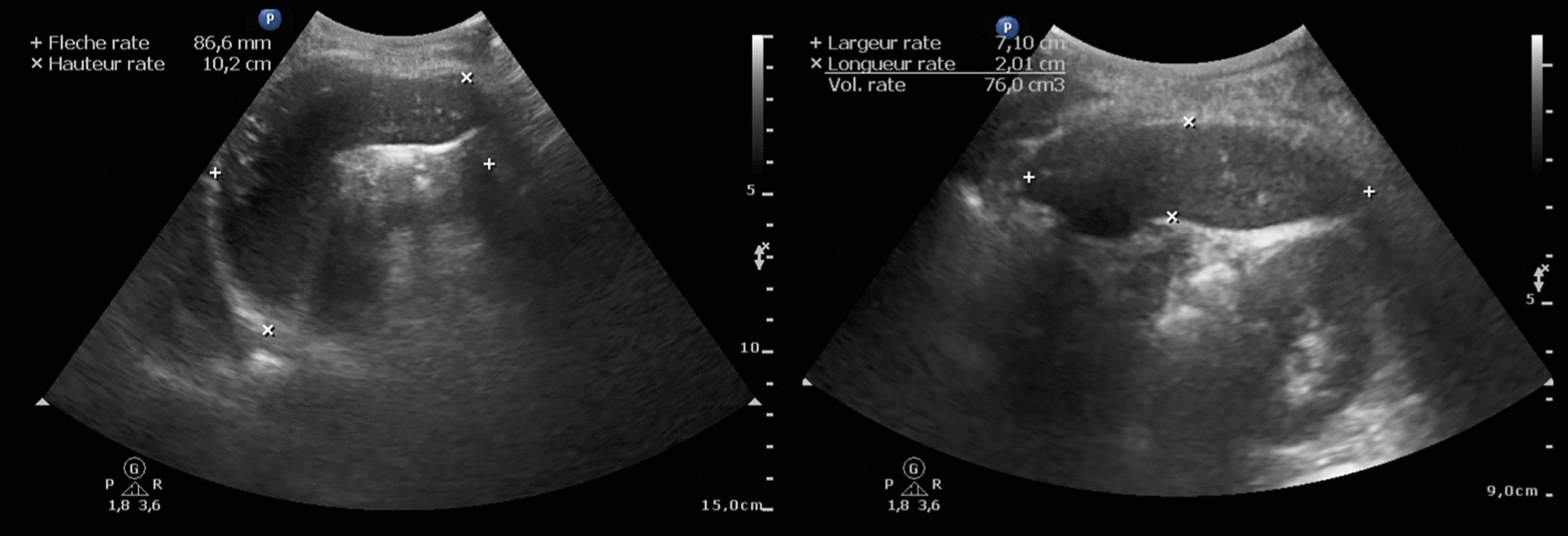
Among the 991 participants enrolled in the parent study, 981 (612 men, 369 women) underwent ultrasound examination. Of these, the spleen could not be visualized in 10 patients, and for them, a zero value was included for all spleen-related measures in the analysis. The socio-demographical and clinical characteristics of the 981 participants are described in Table 1. Splenomegaly (CCD ≥ 13 cm) was observed in 139 out of 981 (14.3%) participants. A total of 26 out of 977 (2.7%), 61 out of 977 (6.2%), and 175 out of 977 (17.9%) individuals had SV ≤ 80, ≤ 110 and ≤ 150 cm3, respectively. Parenchymal lesions on US examination were noted for 42 out of 981 (4.3%) participants, with four of them exhibiting two types of lesions (total number of lesions n = 46). A detailed description of the lesions is given in Table 2. L. loa microfilaremia was found in 353 out of 981 (35.6%) participants, with a median MFD of 2440 mf/ml. One patient had chronic lymphocytic leukemia (SV: 848 cm3; CCD: 11.9 cm), five had circulating lymphoplasmocytes (SV: 258 and 37 cm3; CCD: 11.2 and 5.6) or plasmocytes (SV: 194, 263 and 273 cm3; CCD: 12.1, 9.3 and 9.5 cm). Among participants, there were no cases of hookworm infection or infection with S. mansoni. No schistosomiasis was found among the 94 individuals with hematuria (11 traces/+ and 83 ++ / +++). Only 6 out of 981 individuals (0.6%) had M. perstans mf in their blood (range: 20–660 mf/ml). Twenty-two individuals (2.2%) tested positive for the Ov16 RDT, and none of them had O. volvulus mf in the skin snips.
Table 1 Characteristics of the study populationTable 2 Description of the 46 spleen parenchymal lesions observed in n = 42 patients with spleen lesions on ultrasonographic examinationFactors influencing spleen volume and splenomegaly (Table
3)
Along with age-related atrophy (ß = − 2.17; 95% CI: − 2.86, − 1.48, P < 0.001), SV was larger in males than in females (+ 35.11 cm3; 95% CI: 14.4, 55.82, P = 0.001) and in people with higher BMI (+ 4.59 cm3 per kg·m−2; 95% CI: 1.51, 7.68, P = 0.004); and HJB presence was associated with lower SV (− 26.48 cm3; 95% CI: − 52.03, − 0.93, P = 0.042) (Table 3). Malarial seropositivity titer in IgG was associated with enlarged SV with a gradient effect (global Wald test P < 0.001): + 20.43 (95% CI: − 7.90, 48.76), + 43.63 (95% CI: 13.12, 74.15), + 66.70 (95% CI: 37.07, 96.33) for 50–70, 70–85, and > 85 µg per ml, respectively. Although lacking a significant effect, the Plasmodium PCR had the same tendency. Conversely, the L. loa MFD was associated with lower SV with a gradient effect [Wald global test P = 0.004; − 40.94 cm3 for those with L. loa MFD > 30,000 mf/ml (P = 0.277)], compared to amicrofilaremic ones). Individuals with splenic parenchymal lesions had a mean enlarged SV of 58.23 cm3 (95% CI: 10.14, 106.31, P = 0.018).
Table 3 Factors associated with spleen volume in cm3 or splenomegaly defined as cranio-caudal distance ≥ 13 cm in univariate and multivariable analysisIn the final model, splenomegaly (CCD ≥ 13 cm) was inversely associated with age (aOR = 0.98; 95% CI: 0.97, 0.99, P = 0.004), and associated with the absence of HJB (aOR = 0.4; 95% CI: 0.21, 0.74, P = 0.004). There appeared to be a non-statistically significant association of splenomegaly with the absence of SCD trait (aOR = 0.66; 95% CI: 0.40, 1.09, P = 0.101), and the presence of a lymphopenia (aOR = 1.57; 95% CI: 0.91, 2.68, P = 0.103). Malarial seropositivity was also gradually associated with CCD ≥ 13 cm, with aORs ranging from 1.49 to 3.03 (P = 0.003) for those with the highest serological titers. The reverse was observed with the L. loa MFD (global Wald test P = 0.021): 0.53 (95% CI: 0.30, 0.93), 0.48 (95% CI: 0.22, 1.06), 0.47 (95% CI: 0.20, 1.07), and 0.59 (95% CI: 0.13, 2.70) for 1–2000, 2000–8000, 8000–30,000, and > 30,000 mf/ml, respectively.
Notably, among the patients with CCD ≥ 13 cm, 2 had codocytes, 1 had activated lymphocytes, and 4 had P. falciparum trophozoites.
Among the 139 participants who had spleen CCD ≥ 13 cm, median total serum IgM level (determined for 126 individuals) was 2.2 g/L (interquartile range, IQR: 1.3–3.8). Total IgM > 2.5 g/L combined with CCD ≥ 13 cm (‘possible’ HMS) was seen in 52 participants (5.3%), including 35 males (60.3%); their median age was 56 years (IQR: 45–65). Total IgM levels exceeding the higher threshold (> 3.5 g/L), anti-Plasmodium IgG levels > 85 µg/ml, and CCD ≥ 13 cm (‘probable’ HMS) was combined in 14 participants (1.4%), including 9 males (64.3%); their median age was 57 years (IQR: 45–69). The latter group of 14 participants, exhibited significantly higher lymphocytes levels (Wilcoxon-Mann–Whitney rank test P = 0.04) as compared to controls (median 3300 per µl, IQR: 2500–4500 versus 2700, IQR: 2100–3300). None had hematological malignancy or plasmodial infection on the blood smear. Plasmodium PCR was positive for 19/52 (36.5%) subjects with ‘possible’ HMS and 3/14 (21.4%) subjects with ‘probable’ HMS.
Factors associated with anatomical hyposplenia (Table
4)
Age was positively associated with AH [aORs = 1.05; (95% CI: 1.01, 1.09, P = 0.007), 1.04 (95% CI: 1.02, 1.06, P = 0.001), 1.04 (95% CI: 1.03, 1.06, P < 0.001), for < 80, 110, and 150 cm3, respectively], whereas BMI was negatively associated with AH [aORs = 0.79; (95% CI: 0.67, 0.94, P = 0.007), 0.86 (95% CI: 0.77, 0.95, P = 0.003), 0.91 (95% CI: 0.86, 0.97, P = 0.002), for < 80, 110, and 150 cm3, respectively]. Male sex was negatively associated with AH only in the < 150 cm3 model (aOR = 0.55; 95% CI: 0.38, 0.80, P = 0.001). HJB presence was solely associated with SV ≤ 80 cm3 with aOR of 3.85 (95% CI: 1.55, 9.56, P = 0.004). When considering the HBJ as a categorical variable [0 (reference), ≤ 2%, and > 2%], the final model (for SV ≤ 80 cm3) retained the same variables, with a gradient effect for HBJ (< 2%: aOR = 3.43; 95% CI: 1.21, 9.68, P = 0.020; > 2%: aOR = 4.25, 95% CI: 1.29, 16.63, P = 0.038). A PCR positive Plasmodium infection [aORs = 0.44; 95% CI: 0.18, 1.12, P = 0.086), 0.62 (95% CI: 0.35–1.12, P = 0.116), 0.52 (95% CI: 0.35–0.76, P = 0.001), for < 80, 110, and 150 cm3, respectively], and higher anti-P. falciparum serological levels (> 85 µg per ml) were inversely associated with lower SV [aORs = 0.19; 95% CI: 0.04, 0.94, P = 0.042), 0.52 (95% CI: 0.30, 0.91, P = 0.022), for < 80, and 150 cm3, respectively]. Higher L. loa MFD were associated with increased risk of low SV. A gradient effect was observed in each model, with the highest MFD (> 30,000 mf/ml) having the highest aOR of 17.94 (95% CI: 2.91, 110.76, P = 0.002), 5.94 (95% CI: 1.40, 25.17, P = 0.016), and 5.77 (95% CI: 1.95, 17.12, P = 0.002) for SV ≤ 80, 110, and 150 cm3, respectively.
Table 4 Factors associated with anatomical hyposplenia defined as spleen volume < 80, 110, or 150 cm3 in univariate and multivariable analysisWith L. loa MFD as a binary variable, significant correlations persisted between microfilaremia and reduced SV, employing the same modeling approach as in Table 4. Microfilaremic individuals exhibited adjusted aORs of 2.12 (95% CI: 0.90, 4.99; P = 0.084), 1.82 (95% CI: 1.05, 3.17; P = 0.033), and 1.76 (95% CI: 1.22, 2.54; P = 0.003) for SV ≤ 80 cm3, ≤ 110 cm3, and ≤ 150 cm3, respectively, compared to amicrofilaremic ones.
Codocytes were observed in one patient with SV ≤ 80 cm3, one patient with SV 80–110 cm3, and three patients with SV 110–150 cm3. Conversely, among the 14 patients with codocytes on blood smear, the median SV was 173 cm3 (95% CI: 139, 227, missing data: 1).
Regarding the population attributable fraction (PAF) of SV ≤ 80, ≤ 110, and ≤ 150 cm3 associated with L. loa microfilaremia we found it to be 25.1% (95% CI: 1.5, 43.0), 21.2% (95% CI: 5.5, 34.3), and 18.8% (95% CI: 8.9, 27.6), respectively.
Spleen parenchymal lesions
We observed 8 individuals (0.8%, 95% CI: 0.4, 1.6) presenting with hyperechoic homogeneous macronodules, including 3 cases (0.5%, 95% CI: 0.0, 1.4) among amicrofilaremic individuals and 5 cases (1.4%, 95% CI: 0.5, 3.3) among microfilaremic individuals (P = 0.110). Only 2 hyperechoic micronodules were identified, both of which were found in amicrofilaremic individuals. A total of 8 hypoechoic macronodules (0.8%, 95% CI: 0.4, 1.6) were observed across the population, with 2 cases (0.3%, 95% CI: 0.03, 1.1) among amicrofilaremic individuals and 6 cases (1.7%, 95% CI: 0.6, 3.7) among microfilaremic individuals (P = 0.026). We also identified 12 patients with hyperechoic heterogeneous macronodules or ill-defined hyperechoic lesions, representing 1.2% (95% CI: 0.7, 2.1) of the population. Of these, 7 cases (1.1%, 95% CI: 0.4, 2.3) were observed in amicrofilaremic individuals, and 5 cases (1.4%, 95% CI: 0.5, 3.3) in microfilaremic individuals (P = 0.427). The patient with probable Gamna-Gandy bodies had ascites, compatible with underlying cirrhosis (Table 5).
Table 5 Factors associated with the presence of any spleen lesion in univariate and multivariable analysis 

Comments (0)