
Remember me


A 79-year-old male was admitted to the emergency department at Aalborg University Hospital in Denmark in January 2023 with progressive lower back pain. On admittance, the patient was febrile and noted to have had diarrhoea for at least one week prior to hospitalisation, accompanied by mild complaints of diffuse abdominal pain. The patients’ medical history included prostate cancer, for which he was not yet actively treated, ischemic heart disease, atrial fibrillation, and previous stroke with negligible motor deficits, but the patient was immunocompetent. Vital parameters were stable, but the patient was febrile with a temperature of 38.1°C. Blood tests revealed an elevated C-reactive protein (CRP) level of 227 mg/l, but white blood cell (leucocyte) counts and procalcitonin were within the normal range (< 10 × 10^9/L and < 0.5 µg/L, respectively).
A FecalSwab (Copan Italia, Brescia, Italy) was taken shortly after admittance, which was PCR-positive for Campylobacter spp. A CT scan of the thorax and abdomen showed terminal ileitis and signs of inflammatory discitis at L1/L2 suspect for osteomyelitis. A MRI of columna totalis confirmed suspicion of vertebral osteomyelitis with a hyperintensity signal of the L1/L2 intervertebral disc, and hyperintensity signal of both L1 and L2 vertebral bodies with an epidural abscess, resulting in compression of the dura sack, see Fig. 1. Three days after admission, a blood culture was positive with spiral-shaped, Gram-negative rods, later identified as C. jejuni.
Fig. 1
Sagittal STIR sequence of the lumbar spine showing hyperintensity signal of the L1/L2 intervertebral disc and the vertebral bodies of L1 and L2 with an epidural abscess, resulting in compression of the dura sack (white arrow)
Shortly following the positive blood culture, the patient underwent spinal surgery with spondylodesis and decompression of L1/L2 with donor autolog bonetransplantation in the disc space. The surgical procedure was uncomplicated, and cultivation from the spinal bone retrieved during surgery was also culture-positive for C. jejuni.
On admittance, the patient was empirically started on iv. piperacillin/tazobactam 4 g qad, and upon the blood culture results, iv. clarithromycin 500 mg bid was added. Following spinal surgery, treatment was targeted to iv. meropenem 1 g tid for 4 weeks, followed by 4 weeks treatment with clindamycin (2 weeks at dose 600 mg tid, tapered to 2 weeks dose of 300 mg tid). The patient recovered quickly following surgery, with decreasing lumbar pain and biochemical response, and was fully recovered at three months follow-up in the outpatient clinic. In an attempt to identify the source of C. jejuni infection, the patient reported having eaten undercooked turkey for Christmas dinner one week prior to hospitalization, but no other household members that ate the same meal had become ill.
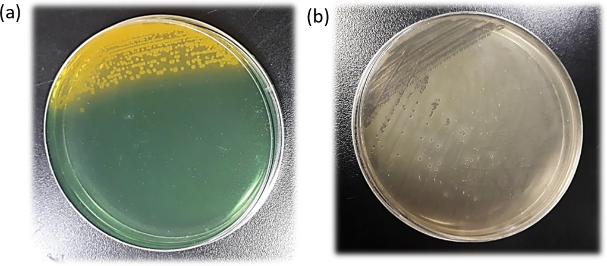
InvestigationsA FecalSwab (COPAN ITALIA, Brescia, Italy) obtained 20 h upon submission was positive for Campylobacter spp. by use of the QIAstat-Dx® Gastrointestinal Panel (Qiagen, Hilden, Germany). A standard blood culture obtained upon submission (Two BD BACTEC™ Plus Aerobic medium and one BD BACTEC™ Lytic Anaerobic medium glass culture vials) was incubated in the BACTEC FX Top instrument (Becton Dickinson AB, Stockholm, Sweden). After three days of incubation there was growth in a single aerobic bottle (Time to detection: 70.4 h) of motile, Gram-negative, spiral-shaped rods. Sub-cultivation was performed on horse blood agar plates (SSI Diagnostica, Hillerød, Denmark) at 37 °C in a hydrogen-enriched microaerobic atmosphere (6% O2, 6% CO2, 6% H2, and 82% N2) and examined the day after (app. 18 h), with growth of non-haemolytic, greyish, smooth, colonies with a swarming growth. Colonies was catalase and oxidase positive and identified as C. jejuni by use of the matrix-assisted laser desorption ionization–time of flight (MALDI Biotyper 3.1, Bruker Daltonics Microflex LT, MBT 6903 MSP Library) with a score of 2.22.
Concurrently, one lumbar bone biopsy specimen (L1-L2) was sent to the microbiological laboratory. The bone specimen was cultured on standard solid culture media and incubated in environments containing 5% CO2, anaerobic conditions, and a hydrogen-enriched microaerobic atmosphere. Additionally, thioglycolate and serum broth were utilized. After 48 h, growth of C. jejuni was observed on all media, including blood agar, which was incubated in environments with 5% CO2, anaerobic conditions, and a hydrogen-enriched microaerobic atmosphere, respectively. Identification was confirmed through high MALDI scores. The bone specimen was also Campylobacter spp. positive by use the standard 16 S rRNA targeted next-generation sequencing platform performed at the reference laboratory at Statens Serum Institut (SSI) [7].
We conducted phenotypic antibiotic susceptibility tests (AST) of C. jejuni following the recommended method outlined in EU Decision 2013/652/EU [8], employing the Sensititre™ EU Surveillance Campylobacter EUCAMP3 AST Plate. For clindamycin, the minimum inhibitory concentration (MIC) was determined using a custom Sensititre™ plate with clindamycin concentration ranging from 0.25 mg/L to 8 mg/L. Following inoculation, the microplates were incubated at 41 °C under microaerobic conditions for 24 h. The results were interpreted according to EUCAST breakpoints (version 13.0) if available, interpreted based on epidemiological cut-off (ECOFF) values, as provided on the EUCAST website (www.eucast.org). The isolate exhibited low minimum inhibitory concentrations (MICs) and were interpreted as sensitive to chloramphenicol (MIC: ≤ 2 mg/L), gentamicin (MIC: ≤ 0.25 mg/L), tetracycline (MIC: ≤ 0.5 mg/L), and erythromycin (MIC: ≤ 1 mg/L), and resistant to ciprofloxacin (MIC: = 8 mg/L). The MIC values for clindamycin and ertapenem were = 0.5 mg/L and ≤ 0.12 mg/L), respectively.
Next, whole-genome sequencing was performed on the Illumina NextSeq instrument using the Nextera XT DNA Library Preparation Kit (Illumina, San Diego, USA) to produce paired-end reads. The raw reads and SKESA assembled genomes were submitted to and passed the SSI in-house QC pipeline (https://github.com/ssi-dk/bifrost). The assemblies were analyzed with the AMRinderPlus (https://github.com/ncbi/amr/wiki), Software version: 3.11.18, Database version: 2023-08-08.2, with settings coverage 0.5, identity 0.9, for the in-silico detection of acquired resistance genes and point mutations. Two resistance mechanisms were detected in the isolate, a point mutation in the gyrA_T86I gene conferring resistance towards ciprofloxacin, and an oxacillinase blaOXA-461 family class D beta-lactamase. The WGS analysis further confirmed the isolate as C. jejuni and by use of the MLST 2.0 webtool (available at: http://www.genomicepidemiology.org) the isolated was assigned to sequence type ST-49.
Comments (0)