
Remember me


 Figure
 Box 1
Box 1Spontaneous coronary artery dissection (SCAD) is a cause of myocardial infarction (MI) not attributable to trauma, atherosclerosis, or iatrogenic causes. SCAD is estimated to account for 1% to 4% of cases of acute coronary syndrome (ACS), but the true prevalence remains obscure, likely because of underdiagnosis.1 SCAD is more common in females, who account for 87% to 95% of cases, with the average presenting age between 44 and 53 years and often with minimal traditional ACS risk factors.2 Specifically, in females under age 50 years, two retrospective, single-site studies in Canada and Japan estimated the prevalence of SCAD in patients with ACS at between 24% and 35%, with the Canadian study showing that 56.9% of females with SCAD were postmenopausal.3,4 The disease is rarer in men, who, if affected, tend to be younger and have SCAD precipitated by activity involving isometric exertion or heavy lifting.5
The cause of SCAD likely is multifactorial, encompassing predisposing arteriopathy and a precipitating stressor. Common predisposing factors for and associated conditions of SCAD include fibromuscular dysplasia (FMD) or other connective tissue disorders, pregnancy and multiparity (four or more births), or a history of hormone therapy.5,6 Systemic inflammatory disease such as lupus erythematous, inflammatory bowel disease, and sarcoidosis also is associated with SCAD, but a recent case-control study suggested that this association is not causative.7 Extravascular abnormalities are common in patients with SCAD, and FMD is the most common of these, with a wide range (25% to 86%) of reported incidence.6
Underlying genetic factors may predispose patients to SCAD, given the low burden of traditional risk factors for coronary disease and higher incidences reported in patients' relatives.2 A 2019 study found that an allele (rs9349379-A) that placed patients at risk for FMD also put them at higher risk for SCAD.8 Certain heritable connective tissue disorders and arteriopathies (Marfan disease, Loeys-Dietz syndrome, Ehlers-Danlos syndrome) are associated with increased risk for SCAD, although these only account for 5% to 9% of cases.2 Despite this, routine genetic screening has not been deemed clinically useful, although patients with a high-risk SCAD phenotype may warrant screening for rare genetic variants.9
 Box 2
Box 2Although many SCAD studies consist mainly of White patients, a racially diverse retrospective cohort study demonstrated similar outcomes and presentations regardless of patient race.10,11 Real-world extrapolation of these results is limited by racial minority underrepresentation in other registries.
PATHOPHYSIOLOGYSCAD occurs when a disruption in the layers of the coronary artery wall creates a false lumen that subsequently fills with an intramural hematoma. Enlarging hematomas can obstruct the true lumen of the coronary artery and induce myocardial ischemia and infarction.12 Two hypotheses are offered for this mechanism: The inside-out hypothesis proposes that the endothelial-intimal layer is disrupted and blood from the true lumen enters to form a hematoma; the outside-in hypothesis contends bleeding occurs in the coronary wall at the level of the vasa vasorum, generating a hematoma without intimal disruption.12 Current evidence favors the outside-in hypothesis, but both likely occur to some degree and may even occur at the same time.
Several structural factors may influence hematoma formation. Observational data using optical coherence tomography (OCT) found that SCAD lesions without fenestration lead to increased pressure in the false lumen, further obstructing the true lumen.13 Vessel tortuosity, defined as three or more consecutive curvatures of 90 to 180 degrees in a major coronary artery of 2 mm or greater in diameter, also has been a prevalent characteristic in SCAD and an associated finding in recurrent cases.14 However, this may be an incidental manifestation of vasculopathy such as FMD.14
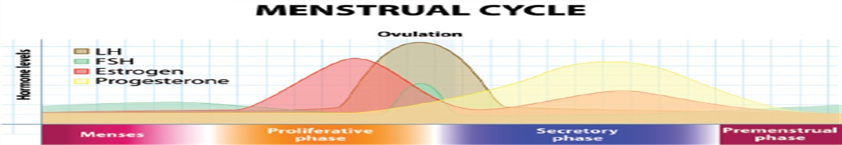
Pregnancy is associated with SCAD (denoted as P-SCAD), and although only 10% of all SCAD cases occur in pregnant or peripartum patients, 21% to 27% of MIs during pregnancy are due to SCAD, and an estimated 50% of postpartum MI events present as SCAD.15 A Canadian study found that P-SCAD cases involved the left main and left anterior descending arteries in 36% and 72% of cases, respectively.16 Multiple artery involvement was found in more than a third of included cases (more than twice the rate of multiartery involvement in other studies).16 Outcomes for patients with P-SCAD often are worse than for nonpregnant patients with SCAD, possibly because of pregnancy-associated changes such as blood volume expansion, hormone-related changes altering the vascular endothelium, and the possible contribution of hormones regulating milk production (several patients described SCAD symptom onset during lactation).17 Although it has been suggested that sex hormones play a role in the development of SCAD (the disease has been reported to occur in patients taking hormone therapy), gaps remain in understanding the interplay of hormones such as estrogen and progesterone in the disease process.2
TYPESSCAD may occur in one or more overlapping conditions identified primarily through angiography (Figure 1).18
Type 1 is diagnosed when contrast dye stains the arterial walls and demonstrates a longitudinal, radiolucent false lumen. Type 2, the most common form, can be misdiagnosed because of the diffuse (often greater than 20 mm), smooth narrowing of the lesion, which can stretch to the distal tip of the artery.2 Type 2 is subdivided into 2a, when the vessel reopens distally as the lesion tapers, and 2b, when the lesion extends the full length of the vessel.12 Type 3 can be difficult to differentiate from atherosclerotic disease because of multiple focal tubular lesions. This type might require intracoronary imaging for confirmation. Type 3 lesions usually are long (11 to 20 mm) and appear hazy and linear on the angiogram.19 Type 4 occurs when intramural hematoma results in a complete distal vessel occlusion.13,20 FIGURE 1.:
 SCAD subtypes on angiogram. (A) Type 1 with radiolucent false lumen. (B) Type 2a in the left anterior descending coronary artery with classic tapering appearance and distal reopening of the vessel. (C) Type 2b with the tapering appearance extending to the end of the vessel. (D) Type 4 with distal vessel occlusion. Type 3 is not pictured because angiographic findings often are difficult to differentiate from atherosclerosis.
CLINICAL PRESENTATION AND DIAGNOSISClinicians should be suspicious of SCAD in younger patients presenting with chest pain. Obtain a thorough history, including a history of a connective tissue disorder such as FMD or a history of hormone therapy. Even greater clinical suspicion should be afforded to pregnant or peripartum patients with this presentation.
As in atherosclerotic ACS, the most common complaint of patients with SCAD is chest pain, reported in up to 96% of cases.21,22 Less frequently, neck and arm pain, dyspnea, nausea, vomiting, diaphoresis, and back pain have been observed; however, few patients have typical ischemic risk factors, initial troponin can be normal (but almost invariably has a minor elevation), and 85% of patients present without ischemic ECG changes.1 Typical risk stratification algorithms such as the History, ECG, Age, Risk Factors, and Troponin (HEART) and the Global Registry of Acute Coronary Events (GRACE) scores may underestimate ACS, although new risk scores for predicting SCAD show promise.23,24 Left ventricular ejection fraction (LVEF) usually is normal (74% to 92.47% of patients have LVEF greater than 50%). Patients with reduced LVEF often normalize, as demonstrated by a prospective Vancouver study showing that 29.9% of patients had reduced LVEF during hospitalization, but that percentage fell to 6.7% during outpatient follow-up at a median of 4.4 months.18,25,26 Up to 10% of patients may present with ventricular dysrhythmias, less than 3% with cardiogenic shock, and less than 1% with sudden cardiac death.18
Presentation and outcomes in patients with P-SCAD are poorer compared with patients with SCAD who are not pregnant.16 Although P-SCAD can occur any time during or after pregnancy, nearly 73% of patients present in the early postpartum period.16 The Mayo Clinic registry found that 57% of patients with P-SCAD presented with ST-segment elevation MI, compared with 36% of patients with SCAD who were not pregnant.17 The incidence of cardiac arrests was nearly equivalent in both groups.17 A case review reported that 16% of patients with P-SCAD presented with ventricular dysrhythmias, compared with 0% to 5% of nonpregnant patients with SCAD in separate studies.16 The same review found that 24% of patients with P-SCAD presented with shock, and maternal mortality was 4%, compared with 0% to 0.5% for both outcomes in nonpregnant patients with SCAD.16
If a workup (troponins, ECG, echocardiogram) is suspicious for ischemia or SCAD, a coronary angiogram is the preferred diagnostic modality. SCAD findings on angiogram include multiple radiolucent lines, contrast staining, appearance of a false lumen, and possible spiral dissection.1 Angiography occasionally can be limited in diagnosis of SCAD because of its lack of sensitivity for arterial wall defects.18 Patients with type 2 or 3 SCAD may need OCT or intravascular ultrasound for better imaging. Intravascular ultrasound can differentiate between atherosclerotic plaque and hematoma, does not require contrast use, and provides the full view of the vessel wall; however, OCT is preferred because of its higher spatial resolution and usefulness in revascularization (it can clarify that the guidewire is in the true lumen for percutaneous coronary intervention [PCI]).15 Coronary CT angiography (CCTA) is not routinely recommended because this imaging technique is limited in diagnostic sensitivity and specificity for SCAD, and current data are based on low-volume studies; however, consider CCTA for patients with known SCAD for follow-up of proximal or large vessel dissections especially if patients have recurrent chest pain.27 CT angiography (CTA) of the neck, chest, abdomen, and pelvis is recommended to rule out FMD because of its significant co-prevalence with SCAD (Figure 2).
 FIGURE 2.:
 (A) Tortuous carotid artery findings indicative of FMD on CTA of the arteries of the neck. (B) Tortuous renal arteries indicating FMD on abdominal CTA.
MANAGEMENTNo randomized controlled trials exist to guide medical management of SCAD, and the optimal approach remains uncertain.15 Most patients with SCAD can be managed conservatively, and expert opinion supports a medical approach in clinically stable patients because of the high rates of spontaneous vessel healing.2,12 A proposed algorithm for SCAD management is shown in Figure 3. Beta-blockers are recommended based on retrospective data showing a lower risk of SCAD recurrence (hazard ratio 0.36 with 95% CI), and this same rationale supports tight BP control.28 Statins are not specifically recommended for patients with SCAD but are prescribed if warranted for primary prevention.12 The optimal duration of dual antiplatelet therapy (DAPT) for patients not undergoing PCI also is unclear, but evidence from the DIssezioni Spontanee COronariche (DISCO) registry indicates that single antiplatelet therapy is the preferred strategy for conservatively managed patients, because solo aspirin is associated with a lower risk of adverse reactions.2,29 Obtain an echocardiogram during admission, regardless of patient stability, to guide medical care and the potential for device therapy. The American Heart Association heart failure guidelines can be followed, as applicable, when treating these patients.30 If the patient's LVEF is reduced at the time of presentation, reassess cardiac function via an echocardiogram about 3 months after presentation.2
 FIGURE 3.:
 A proposed algorithm for approaching patients with confirmed SCAD
Patients with hemodynamic instability, ongoing ischemia, or an unstable ventricular dysrhythmia may require PCI; however, this approach should be attempted with caution because of the potential for iatrogenic dissection and low success rates.1,28 If PCI is performed, guidelines can be followed for DAPT afterwards as with patients with other forms of ACS undergoing PCI; whether the duration of therapy should differ in patients with SCAD compared with those with atherosclerosis is unclear.2,31 Patients with high-risk anatomy (severe dissection in the proximal portion of two or more vessels or left main coronary artery involvement) or those in whom PCI was unsuccessful may require coronary bypass grafting (CABG).12,15 Contemporary reviews note that much of the positive data regarding this approach is limited to small-case series with high rates of late graft occlusion likely due to competitive flow from the native coronary artery as the dissection heals.12,15 Even with high rates of graft occlusion, long-term mortality is comparable to conservative management, and high-risk patients should not be excluded from CABG if it may benefit them.2
Pregnant patients should avoid ionizing radiation and drugs harmful to the fetus (if a beta-blocker is prescribed, labetalol is recommended).1,15 A conservative approach is preferred in patients without high-risk features, with care tailored by a multidisciplinary team including an obstetrician if available. If the fetus is at a viable age and timing of delivery is in question, scenarios that may be appropriate for proceeding with delivery include nonreassuring fetal heart tracings, the mother suffering cardiac arrest, or possible consideration of delivery before planned CABG.32
Conservatively managed patients should be monitored while inpatient for 3 to 5 days because of the high rates of recurrent chest pain and potential for hematoma extension, and ultimately, Hayes and colleagues recommend that length of stay should be individualized and err on the longer side, compared with patients with atherosclerotic ACS.2
PROGNOSIS AND RECURRENCEThe European Society of Cardiology writing committee noted that SCAD recurrence rates vary widely, from 4.7% to 29.4% depending on timing of follow-up, definition of recurrence used, and the study design.15 Adlam and colleagues also documented low long-term mortality, with survival rates ranging from 92% to 100%, but 14.6% to 47.4% of patients had major adverse cardiac events, mainly driven by recurrent dissections and PCI failures.15
Timing of recurrence varies but typically happens soon after initial discharge, and patients should be warned about symptoms that could indicate hematoma or dissection flap extension.2 Patients may continue to experience intermittent chest pain after hospital discharge, requiring another admission. These patients should be assessed with high-sensitivity troponin and ECG.1 Because of concerns about secondary iatrogenic dissection if an interventional approach is attempted, angiography should be reserved for patients with significant concern for high-risk ischemia or abnormal functional tests.1 Unfortunately, no therapy has been proven to reduce SCAD recurrence rates at this time. Beta-blocker therapy and good BP control offer some promise, but these approaches require further study.28
Because of the association of SCAD and pregnancy, patients should be educated on family planning and contraceptive methods that can help avoid recurrent pregnancy following SCAD, although most patients who become pregnant do not suffer a recurrence of SCAD.2,33
OTHER CONSIDERATIONSLike patients with other forms of ACS, those with SCAD should be referred to cardiac rehabilitation after hospital discharge.1 Cardiac rehabilitation generally is covered by insurance if a guideline-directed qualifying diagnosis is made.31 Programs should be tailored to the patient, considering factors such as age, baseline ability to exercise, and LVEF. No strong studies have assessed the outcomes of weight and aerobic restrictions for physical activity in patients with SCAD. Until more robust studies are completed, clinicians should follow the Vancouver Hospital program model, which recommends 50% to 70% of the heart rate reserve and free weight use of no more than 12 lb (5.4 kg), with gradual up-titration.34 Long-term recommendations also should be individualized, but patients should avoid intense endurance training, exercise in the extremes of temperature, and high-level competitive sports.2
CONCLUSIONSCAD is an underdiagnosed cause of ACS, especially in younger women and peripartum patients. SCAD presents as several different types, some of which create the opportunity for misdiagnosis. Suspect SCAD in younger patients with typical ACS symptoms, especially pregnant or postpartum patients and those who have recently had strenuous physical activity (particularly isometric exertion or heavy lifting). Most conservatively managed patients have good long-term survival, but recurrent symptoms of varying severity are frequent. Interventional and even surgical approaches may be needed for high-risk patients. Until more robust studies are generated, patients with SCAD should avoid extreme physical exertion, maintain BP control, and discuss the risks and benefits of any hormone therapy and future pregnancy planning with an expert cardiology team.
REFERENCES 1. Hayes SN, Kim ESH, Saw J, et al. Spontaneous coronary artery dissection: current state of the science: a scientific statement from the American Heart Association. Circulation. 2018;137(19):e523–e557. 2. Hayes SN, Tweet MS, Adlam D, et al. Spontaneous coronary artery dissection: JACC state-of-the-art review. J Am Coll Cardiol. 2020;76(8):961–984. 3. Meng P-N, Xu C, You W, et al. Spontaneous coronary artery dissection as a cause of acute myocardial infarction in young female population: a single-center study. Chin Med J (Engl). 2017;130(13):1534–1539. 4. Saw J, Aymong E, Mancini GB, et al. Nonatherosclerotic coronary artery disease in young women. Can J Cardiol. 2014;30(7):814–819. 5. Fahmy P, Prakash R, Starovoytov A, et al. Pre-disposing and precipitating factors in men with spontaneous coronary artery dissection. JACC Cardiovasc Interv. 2016;9(8):866–868. 6. Janssen EB, de Leeuw PW, Maas AH. Spontaneous coronary artery dissections and associated predisposing factors: a narrative review. Neth Heart J. 2019;27(5):246–251. 7. Kronzer VL, Tarabochia AD, Lobo Romero AS, et al. Lack of association of spontaneous coronary artery dissection with autoimmune disease. J Am Coll Cardiol. 2020;76(19):2226–2234. 8. Adlam D, Olson TM, Combaret N, et al. Association of the PHACTR1/EDN1 genetic locus with spontaneous coronary artery dissection. J Am Coll Cardiol. 2019;73(1):58–66. 9. Wang Y, Starovoytov A, Murad AM, et al. Burden of rare genetic variants in spontaneous coronary artery dissection with high-risk features. JAMA Cardiol. 2022;7(10):1045–1055. 10. Clare R, Duan L, Phan D, et al. Characteristics and clinical outcomes of patients with spontaneous coronary artery dissection. J Am Heart Assoc. 2019;8(10):e012570. 11. Chen S, Merchant M, Mahrer KN, et al. Spontaneous coronary artery dissection: clinical characteristics, management, and outcomes in a racially and ethnically diverse community-based cohort. Perm J. 2019;23:18.278. 12. Garcia-Guimarães M, Bastante T, Antuña P, et al. Spontaneous coronary artery dissection: mechanisms, diagnosis and management. Eur Cardiol. 2020;15:1–8. 13. Jackson R, Al-Hussaini A, Joseph S, et al. Spontaneous coronary artery dissection: pathophysiological insights from optical coherence tomography. JACC Cardiovasc Imaging. 2019;12(12):2475–2488. 14. Eleid MF, Guddeti RR, Tweet MS, et al. Coronary artery tortuosity in spontaneous coronary artery dissection: angiographic characteristics and clinical implications. Circ Cardiovasc Interv. 2014;7(5):656–662. 15. Adlam D, Alfonso F, Maas A, et al. European Society of Cardiology, Acute Cardiovascular Care Association, SCAD study group: a position paper on spontaneous coronary artery dissection. Eur Heart J. 2018;39(36):3353–3368. 16. Havakuk O, Goland S, Mehra A, Elkayam U. Pregnancy and the risk of spontaneous coronary artery dissection: an analysis of 120 contemporary cases. Circ Cardiovasc Interv. 2017;10(3):e004941. 17. Tweet MS, Hayes SN, Codsi E, et al. Spontaneous coronary artery dissection associated with pregnancy. J Am Coll Cardiol. 2017;70(4):426–435. 18. Saw J, Mancini GB, Humphries KH. Contemporary review on spontaneous coronary artery dissection. J Am Coll Cardiol. 2016;68(3):297–312. 19. Yip A, Saw J. Spontaneous coronary artery dissection—a review. Cardiovasc Diagn Ther. 2015;5(1):37–48. 20. Mori R, Macaya F, Giacobbe F, et al. Clinical and angiographic features of SCAD type 4. Int J Cardiol. 2023;377:22–25. 21. Luong C, Starovoytov A, Heydari M, et al. Clinical presentation of patients with spontaneous coronary artery dissection. Catheter Cardiovasc Interv. 2017;89(7):1149–1154. 22. Lindor RA, Tweet MS, Goyal KA, et al. Emergency department presentation of patients with spontaneous coronary artery dissection. J Emerg Med. 2017;52(3):286–291. 23. Huang Z, Wang K, Yang D, et al. The predictive value of the HEART and GRACE scores for major adverse cardiac events in patients with acute chest pain. Intern Emerg Med. 2021;16(1):193–200. 24. Shafi I, Maruthi R, Khalid MU, et al. Derivation and validation of spontaneous coronary artery dissection prediction score in patients with myocardial infarction. Am J Cardiol. 2023;201:170–176. 25. Zhao XY, Li JX, Tang XF, et al. Prognostic value of NT-proBNP in stable coronary artery disease in Chinese patients after percutaneous coronary intervention in the drug-eluting stent era. Biomed Environ Sci. 2018;31(12):859–866. 26. Franco C, Starovoytov A, Heydari M, et al. Changes in left ventricular function after spontaneous coronary artery dissection. Clin Cardiol. 2017;40(3):149–154. 27. Pozo-Osinalde E, García-Guimaraes M, Bastante T, et al. Characteristic findings of acute spontaneous coronary artery dissection by cardiac computed tomography. Coron Artery Dis. 2020;31(3):293–299. 28. Saw J, Humphries K, Aymong E, et al. Spontaneous coronary artery dissection: clinical outcomes and risk of recurrence. J Am Coll Cardiol. 2017;70(9):1148–1158. 29. Cerrato E, Giacobbe F, Quadri G, et al. DISCO Collaborators. Antiplatelet therapy in patients with conservatively managed spontaneous coronary artery dissection from the multicentre DISCO registry. Eur Heart J. 2021;42(33):3161–3171. 30. Heidenreich PA, Bozkurt B, Aguilar D, et al. 2022 AHA/ACC/HFSA guideline for the management of heart failure: a report of the American College of Cardiology/American Heart Association Joint Committee on Clinical Practice Guidelines. Circulation. 2022;145(18):e895–e1032. 31. Amsterdam EA, Wenger NK, Brindis RG, et al. 2014 AHA/ACC guideline for the management of patients with non-ST-elevation acute coronary syndromes: executive summary: a report of the American College of Cardiology/American Heart Association Task Force on Practice Guidelines. Circulation. 2014;130(25):2354–2394. 32. Codsi E, Tweet MS, Rose CH, et al. Spontaneous coronary artery dissection in pregnancy: what every obstetrician should know. Obstet Gynecol. 2016;128(4):731–738. 33. Tweet MS, Young KA, Best PJM, et al. Association of pregnancy with recurrence of spontaneous coronary artery dissection among women with prior coronary artery dissection. JAMA Netw Open. 2020;3(9):e2018170. 34. Chou AY, Prakash R, Rajala J, et al. The first dedicated cardiac rehabilitation program for patients with spontaneous coronary artery dissection: description and initial results. Can J Cardiol. 2016;32(4):554–560.
Comments (0)