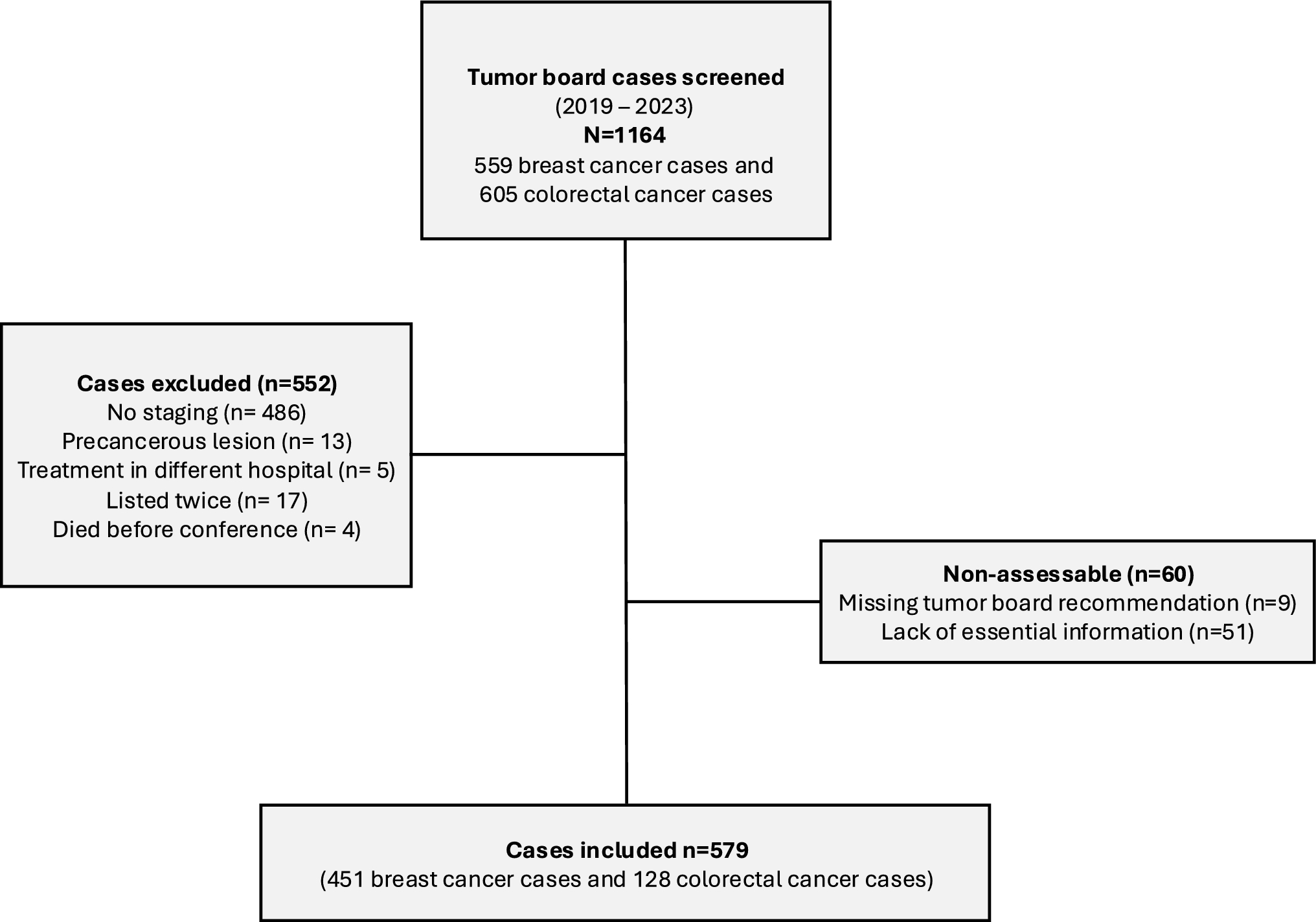
Datasets and source
This study utilized diverse datasets obtained from publicly available databases to investigate various aspects of cancer. The TCGA pancancer cohort provided valuable resources, including mRNA expression, copy number variation, masked copy number partitioning, and DNA methylation 450 K data. These datasets were accessed through the Firehose database, which offers unrestricted access to both tumor and normal samples (http://gdac.broadinstitute.org). Additionally, the UCSC Xena database was utilized to acquire miRNA, TCPA, mutation data, molecular subtyping, and clinical data (https://xenabrowser.net/datapages/). The GEO database was used for validating external mRNA-level data (https://www.ncbi.nlm.nih.gov/geo/), while the CPTAC database contains mass spectrometry data at the protein level (https://proteomics.cancer.gov/programs/cptac). Immunohistochemistry and immunofluorescence data were obtained from the HPA database. For the evaluation of the pancancer immune infiltration results, the TIMER 2.0 database provided multiple immune infiltration algorithms (http://timer.cistrome.org/).
It is important to highlight that these aforementioned public databases are freely accessible, and the study strictly adhered to the policies set by each database during the data extraction process. Consequently, no ethical review or approval was necessary.
Multiomics differential expression analysis
The primary objective of this investigation was to explore the dysregulation of DKC1 gene expression in tumors and normal tissues and to derive correlation results via multidimensional difference analysis. Due to the insufficient number of normal tissue samples in the TCGA dataset, we integrated data from the GTEx to increase confidence and sample size. We used the Wilcoxon test for difference detection, and the significance level was set at p < 0.05. To better understand the distribution of DKC1 gene expression in different organs, we used the “gganatogram” package for visualization analysis. Next, in this study, a comparison was made to assess the differences in DKC1 mRNA expression between tumor tissues and adjacent normal tissues in TCGA, and a Wilcoxon test analysis was performed on paired samples in the TCGA cancer subgroup. To assess the importance of the DKC1 gene in a wide range of cancer diagnostics, we used the “pROC” package to calculate the area under the curve (AUC) values.
External validation and immunohistochemistry in clinical tissues
In this study, we used multiple validation methods. We used the GEO database to validate the accuracy of the gene expression data. To ensure the validity and accuracy of the findings, we also used CPATAC-based mass spectrometry data and immunohistochemistry data from the HPA database to validate protein expression.
SCNA, mutation and DNA methylation analysis
cBioPortal is a comprehensive database for cancer genomics-related data that provides various querying and visualization tools for multiple types of genomic data, including somatic mutation, DNA copy number alteration (CNA), and DNA methylation data (Cerami et al. 2012). In the analysis of copy number alterations (CNAs) and mutations, we employed heterozygosity and homozygosity states to determine the copy number variation of each gene, defining genes with alterations in more than 5% of the samples as high-frequency CNAs. Spearman correlation was utilized to assess the relationship between CNAs and the expression of the DKC1 gene, and the degree of association between gene expression values and copy number segment values was calculated.
Furthermore, we used the R package “IlluminaHumanMmethylation-450kanno.ilmn12. hg19” from Bioconductor to annotate the methylation status of each gene’s promoter. Through the Wilcoxon rank sum test, we performed differential methylation testing on each gene in tumor samples and normal samples to identify genes exhibiting significantly lower or higher methylation. Finally, we computed the Spearman correlation between gene transcription and promoter DNA methylation beta values. To identify significant correlations, a p value threshold of less than 0.05 was adopted as the statistical criterion in this study. We utilize cBioPortal to select the “TCGA Pan Cancer Atlas Study”12 and input the DKC1 gene for a feature search to evaluate the association between copy number alterations (CNAs) and mutations in the DKC1 gene and its expression.
TME and validation through single-cell analysis
The TME exerts a fundamental influence on the progression and pathogenesis of malignant tumors, with interactions between tumor cells being of utmost importance. To gain deeper insights into these interactions, a comprehensive investigation was undertaken to examine the potential associations between the expression of DKC1 and genes related to immune responses. In this study, we calculated the TIP score and subsequently conducted a comparative analysis of the differences in TIP between two distinct groups categorized based on the expression levels of the DKC1 gene. To assess the significance of the observed differences, we employed the Wilcoxon test. For a more accurate assessment of immune infiltration levels, we utilized the TIMER2.0 platform and analyzed the TCGA tumor spectrum using seven advanced algorithms, namely, TIMER, XCELL, MCPCOUNTER, QUANTISEQ, EPIC, CIBERSORT, and CIBERSORT_ABS. Furthermore, we validated the results of the TME by leveraging single-cell datasets from the TISCH database and obtained gene expression profiles across different single-cell datasets. In summary, we performed a comprehensive analysis and visualization of a pancancer cohort to explore the relationship between the TME and gene immune infiltration.
Pathway and functional mechanism analysis
To investigate the relationships between genes and different types of tumors, a series of analyses were conducted. First, based on the DKC1 expression levels, the tumor samples were stratified into two distinct groups. Then, GSEA was employed to explore the activity of gene sets within these groups, including the activation or inhibition status of 50 hallmark gene sets and 74 metabolism gene sets. Additionally, single-cell analysis data were processed using the CancerSEA (Yuan et al. 2019) website, and 14 functional states were redefined. The z score algorithm was utilized to calculate the gene set activity for these functional states, reflecting the pathway activity of interest, and the correlation between genes and functional states was evaluated through pearson correlation analysis. Furthermore, the relationships between the six cell death scores and gene mRNA expression levels were investigated. To gain insight into the potential impact of DKC1, a comprehensive analysis utilizing differential KEGG enrichment was conducted to elucidate the specific pathways that they may modulate. Protein‒protein interaction data were screened, and proteins interacting with DKC1 were determined using protein localization and interaction scores (Veres et al. 2015). Finally, gene-related proteins were identified using CRISPR knockout screen-derived gene effect scores, and the associations between genes and proteins were systematically studied using Spearman correlation analysis.
Analysis of clinical variables and molecular subtypes
To explore the differences in the gene expression levels of DKC1 across diverse tumor subtypes, we used the Kruskal‒Wallis test. Additionally, we used the chi-square test as a statistical method to compare and evaluate differences in molecular subtypes and clinical characteristics between groups with high and low DKC1 expression. Furthermore, we accessed comprehensive cancer immunosubtype information from the USCS Xena database, encompassing wound healing type (C1), IFN-γ-dominant type (C2), inflammatory type (C3), lymphocyte-depleted type (C4), immune quiescent type (C5), and TGF-β-dominant type (C6). These subtypes provided detailed descriptions of the tumor’s immune status. We assessed the variations in DKC1 expression across diverse molecular subtypes of tumors and subsequently investigated the associations between groups exhibiting divergent levels of DKC1 expression in relation to molecular subtypes and clinical characteristics.
Survival and clinical outcome analysis
This study investigated the relationships between DKC1 gene expression and several prognostic indicators, including disease-specific survival (DSS), overall survival (OS), the progression-free interval (PFI), and the disease-free interval (DFI). And we searched the survival data using the TCGA database, applied the “survival” and “survminer” packages in R, and used two statistical methods, namely, the Kaplan‒Meier method and one-way Cox analysis, to comprehensively evaluate the relationship between DKC1 and tumors. In the Kaplan‒Meier survival analysis, we determined the optimal cutoff values for the different DKC1 mRNA expression groups using the “survminer” package and performed a log-rank test using the Surfeit function to assess significant differences. In addition, we used the “forestplot” package to visualize the results of the Cox analysis.
Identification of chemical compounds that interact with DKC1
The Comparative Toxicogenomics Database (CTD) (Davis et al. 2021) provides toxicological information on chemical substances, genes, phenotypes, diseases, and exposures, which helps us gain a deeper understanding of their potential impacts on health. Additionally, we performed gene expression and drug sensitivity analyses using the GSCALite database (Liu et al. 2023). This database includes information on 750 small-molecule drugs and combines it with gene expression data to reveal valuable insights into the associations between these drugs and DKC1. To further explore the relationship between drugs and DKC1 expression, we used a cancer cell line platform created by the National Cancer Institute (NCI) of the United States, which includes 60 human cancer cell lines covering nine different types of cancer. By analyzing the DKC1 expression data and drug sensitivity scores of these cell lines, we calculated Spearman correlation coefficients and identified genes whose expression significantly differed among various cancer types. From these findings, we selected 150 cancer-related genes as markers and conducted matching score calculations using the CMAP_gene_signatures. RData file (Malta et al. 2018; Yang et al. 2022). Finally, we summarized and visualized the research results for 32 cancer types using the R programming language.
Colony formation assay
For the colony formation assay, EC9706 and KYSE30 cells were transfected with negative control and ShDKC1 (2 × 10² cells/well) in a 6-well plate (Corning, NY, USA) and cultured for two weeks. Afterward, the colonies were stained with 0.01% crystal violet for 10 min.
Transwell migration assay
For the Transwell migration assay, EC9706 and KYSE30 cells were plated into the upper chambers of Transwell units (pore size, 8 μm, Corning). The lower chamber was filled with DMEM medium supplemented with 20% FBS. The Transwell units were incubated for 36 h and stained for 5 min. Cell migration onto the lower surface was quantified through microscopic examination.
Cell counting Kit-8 (CCK8) assay
Logarithmic growth phase ESCA cells EC9706 and KYSE30 were seeded into a 96-well plate. After cell adhesion, the culture medium containing different concentrations (0, 20, 100, 200, 400, 800 μM) of Fasudil was added to the esophageal cancer cells and incubated for 48 h. Then, the medium containing the drug was carefully aspirated along the walls of the 96-well plate, and 100 μL of freshly prepared medium containing 10% CCK-8 was added to each well. The plate was placed in a cell culture incubator at 37 °C for 2 h while avoiding light exposure. The absorbance (OD) value at 450 nm was measured using a microplate reader, and the half-maximal inhibitory concentration (IC50) of Fasudil was calculated.
RNA collection and qPCR
The clinical samples in this study of biological tissue originated from the First Affiliated Hospital of Zhengzhou University. This study has been approved by the Ethics Committee of Zhengzhou University. Tissue RNA Purification Kit Plus (EScience Biotech, Shanghai, China) was used to obtain RNA samples from nine ESCA tissue cases and eight adjacent tissue cases as the manufacturer’s instructions. Subsequently, the RNA was converted into cDNA using the UEIris qPCR System for First-Strand cDNA Synthesis. For qPCR amplification, the Applied Biosystems QuantStudio5 (Thermo Fisher Scientific, MA, USA) was used in a 20 μL reaction volume, which included 10 μL of Universal SYBR Green qPCR Supermix (US Everbright, Suzhou, China). The primers were synthesized by Sangon Biotech (Shanghai, China), and PCR was repeated three times for validation. The mRNA expression levels were normalized to GAPDH expression values. The utilized primer pairs were as follows: GAPDH: 5′-ATCACCATCTTCCAGGAGCGAG-3′ (forward), 5′-CATTGCTGATGATCTTGAGGCTGT-3′ (reverse); DKC1: 5′-TAACATGGCGGATGCGGAAG-3′ (forward), 5′-TCAGCGTGTTGTATTTCGGCTTGT-3′ (reverse).
Immunohistochemistry (IHC)
DKC1 antibody from Proteintech (Cat. 25420–1-AP, Wuhan, China) was used for immunohistochemistry on 27 ESCA tissue samples and 29 corresponding adjacent normal tissue samples obtained from the First Affiliated Hospital of Zhengzhou University. SPlink Detection Kits (Origene, Beijing, China) were used for IHC of DKC1(diluted at 1:500). An EDTA-based pH 9.0 solution was used for retrieval. The images were captured using a Zeiss Axio Scope 5 microscope.
Tumor xenograft model
BABL/c male nude mice aged 4–6 weeks were obtained from Gempharmatech Company (China) and housed in isolator cages under specific pathogen-free (SPF) conditions. The animal procedures adhered to the guidelines endorsed by the Institutional Animal Care and Use Committee of Zhengzhou University, ensuring ethical conduct and compliance (Approval NO. ZZU-LAC2020111707). Mice were provided with a sterilized chow diet and water ad libitum. A total of 1.5 × 10⁶ stably transfected vector control or EC9706 ShDKC1 cells were subcutaneously injected into the mice. Tumor volumes were measured twice a week.
Statistical analysis
Statistical analysis was conducted using GraphPad Prism 7 software with Student's t test and one-way ANOVA.


Comments (0)